
FRCEM SBA image quality blurry x-ray problem
TL;DR — Blurry x-rays, ECGs and clinical photos in the FRCEM SBA are a recurring, real candidate complaint, not a personal failing. On the day: use the image viewer’s zoom, treat soft images as a clinical-reasoning question rather than a pixel-counting exercise, and flag the question via the in-test review feature. Within 24 hours of leaving the centre, email exams@rcem.ac.uk with the question reference if you can recall it. Appeals only succeed for procedural irregularity, so document everything in real time.
Facts last verified: 30 May 2026
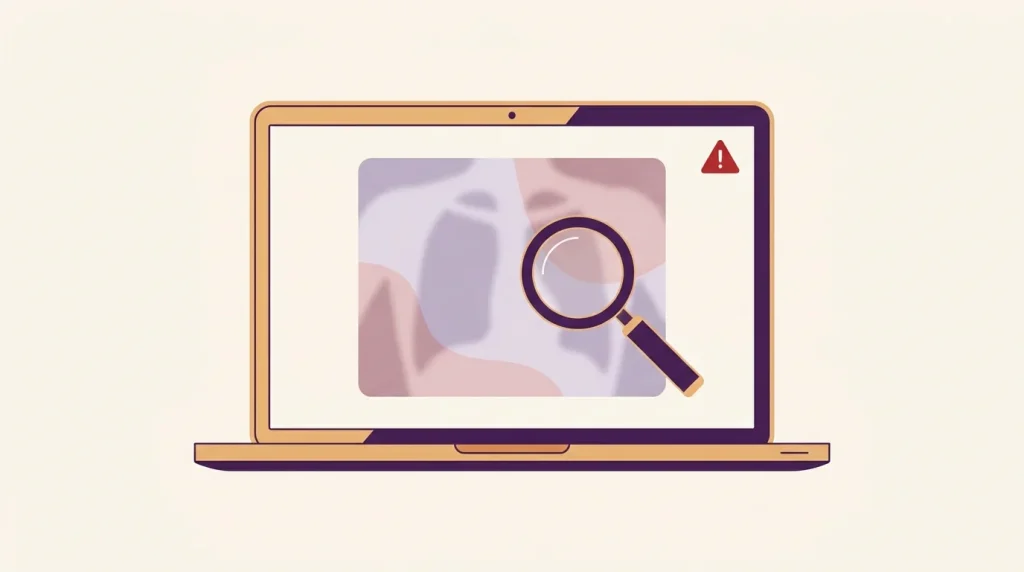
If you came out of an FRCEM SBA sitting convinced that several of the x-rays or ECGs looked low-resolution, washed out or off-colour on the test-centre screen, you are not imagining it and you are not alone. The complaint surfaces after most diets on r/MRCEM and r/JuniorDoctorsUK, and the College’s published position quietly concedes that image quality varies between questions while maintaining that every image is "of sufficient quality" to reach the correct answer. The honest position for candidates is somewhere in between: the images are usually just about workable, but the burden of compensating for soft rendering is shifted onto you. This article validates the complaint, explains the likely technical reasons, gives you a practical playbook for the moment it happens, and walks through the feedback and appeals routes if you think a specific question was unfair.
For more on this, see our guide to RCEM exam centre locations UK.
Is the FRCEM SBA blurry images complaint real or just exam-day stress?
It is real. The pattern recurs in Reddit threads on r/MRCEM and r/JuniorDoctorsUK after almost every diet — candidates describe x-rays that look like low-resolution JPEGs, ECGs where the small squares blur into each other on standard zoom, and dermatology photos where colour balance makes diagnosis ambiguous. We are characterising this sentiment from publicly visible thread context rather than quoting individuals, because the threads themselves are not always fully accessible to external tools, but the volume and consistency of the complaint over time is what matters. The College itself has acknowledged in writing that "images used in our exams have been reviewed and selected by our lead examiners, whilst some may be of higher quality than others, all have been deemed of sufficient quality to help determine the correct answer." That sentence does not say image quality is uniform. It says variability is expected and considered tolerable.
What that means for you, sitting at a Surpass test centre or an OnVUE-style remote sitting: do not assume you have misread the image because it looks worse than what you have practised on. The practice question banks render on a modern personal monitor or laptop with sharp source images. The exam renders compressed images on a fixed test-centre setup. Some loss is structural.
Why do x-rays look blurry on the FRCEM SBA?
There are three plausible reasons, and most candidates encounter a mixture of all three in a single sitting.
- Source image compression. Exam question images are usually delivered as JPEGs sized to a fixed dimension. JPEG compression destroys high-frequency detail — fine lines on an ECG, the trabecular pattern of bone, subtle parenchymal markings on a chest film. The smaller the file and the higher the compression ratio, the more is lost.
- Test-centre monitor specifications. Pearson VUE’s published professional test centre requirements specify 24-inch monitors at 1920×1080 resolution. Surpass test centres also publish a 1920×1080 minimum. That is a perfectly adequate office display, but it is well short of the 4K or medical-grade monitors most candidates use to review images clinically. A chest x-ray that fills a 1920×1080 viewport is being shown at a fraction of the pixel density a PACS workstation would use.
- Scaling and colour calibration. Test-centre monitors are not calibrated for medical imaging. Greys are not reliably greys; subtle density gradients on a film can flatten or distort. Browser-based assessment platforms also rescale images to fit the viewport, which softens edges further.
None of this is unique to RCEM. Every UK and international postgraduate exam that uses computer-based delivery confronts the same problem. What feels frustrating about the FRCEM SBA specifically is that emergency medicine candidates are routinely tested on imaging interpretation in their daily work on PACS, where pan, zoom, window, level and side-by-side comparison are all standard. On a test-centre monitor, most of that goes away.
For more on this, see our guide to Pearson VUE & Surpass test centre noise playbook.
Is the FRCEM SBA still delivered by Pearson VUE in 2026?
No — and this matters for image rendering. From January 2026 RCEM’s theory exams (MRCEM Primary, MRCEM SBA and FRCEM SBA) moved from Pearson VUE to Surpass Assessment as the delivery partner. Surpass operates both a test-centre network and an online-invigilation route. The relevant feature for our purposes is that Surpass markets a high-fidelity image viewer for assessments containing radiographs and similar media, which on paper allows candidates to zoom into images. Whether the FRCEM SBA build of the platform exposes that viewer to every image, or only to a subset, is something candidates from the first 2026 diets are still characterising in real time.
Practical implication: if you sat the exam before January 2026, you saw Pearson VUE’s static image rendering. If you sat after, you are on Surpass, which may give you more interactive control over images. Look for a magnifier or zoom icon on each image-bearing question and try clicking it early in the paper so you know it works before you need it under pressure.
For more on this, see our guide to FRCEM SBA flagging strategy.
Can I zoom into a blurry x-ray during the exam?
Almost certainly, yes, but the controls are not always obvious. On the Surpass platform, the image viewer for radiographs and similar media supports zoom and pan as a documented feature of the platform. Practical steps the moment you hit a blurry image:
- Click directly on the image. On most builds this opens a larger or zoomable view.
- Look for a magnifier, plus-symbol or fullscreen icon adjacent to the image thumbnail.
- Try the browser’s own keyboard zoom (Ctrl with + / −) only if the question is genuinely unreadable and you have already tried the in-platform controls. Some platforms disable this; it should not be relied on, and if you use it you must remember to restore the original zoom before submitting so layout does not break.
- If you can move closer to the screen comfortably, do — moving from arm’s length to about 40 cm doubles the angular size of the detail you are trying to see.
Do not waste two minutes hunting for a zoom that does not exist. If thirty seconds of trying the obvious controls has not improved the image, accept the rendering you have and shift to clinical reasoning.
Can I ask the invigilator to help with a blurry image?
You can raise your hand, and you should if you genuinely believe there is a technical fault — a frozen image, a question that has failed to load an image at all, a completely black or corrupted thumbnail. Invigilators can log the issue and in some cases call Surpass technical support. What they will not do is interpret the image with you, adjust the platform’s zoom on your behalf in any meaningful way, or substitute the question. Their remit is technical integrity of the sitting, not exam content.
If you do raise a technical issue, get the invigilator to record the question number, the time, and a brief description of the fault in the centre’s incident log. Ask whether an incident report is being submitted to RCEM. This is the documentation you will need if you later want to give feedback or, in extreme cases, appeal on grounds of procedural irregularity.
How should I answer an SBA question when the image is genuinely unreadable?
Treat it as a clinical-reasoning question rather than an image-interpretation question. The stem almost always contains enough discriminator information to narrow the answer even without a clean image. Apply the standard SBA priorities — read the lead-in first, classify the task, assess stability, generate a short differential from the history and observations alone, and only then go back to the image to confirm or refute. Several FRCEM SBAs that look like x-ray questions are really pattern-recognition questions where the radiograph confirms what the history already suggests.
Concrete examples of what you can fall back on:
- Chest x-ray you can’t read clearly: a young patient with sudden pleuritic chest pain, reduced breath sounds on one side, hyper-resonant percussion — the stem already points at pneumothorax. The image is confirmatory, not the entire diagnostic step.
- ECG with blurred small squares: rate and rhythm can usually still be estimated. Look for ST elevation, broad versus narrow complexes, and obvious AV dissociation — these survive low resolution far better than P-wave morphology.
- Dermatology photo with poor colour: rely on history (timeline, distribution, contacts, systemic features) and look at distribution and shape rather than colour saturation.
- CT slice that looks soft: the answer is usually about which anatomical compartment is involved (extradural vs subdural, posterior vs anterior circulation infarct, free air vs free fluid) rather than fine texture.
Flag the question for review using the in-platform flag or review function, answer your best clinical guess so you have something committed, and move on. Return at the end with whatever time remains.
Should I bring my own glasses or contact lenses?
Yes, and this matters more than candidates expect. Test-centre monitors sit at a fixed distance and ambient lighting is usually high to reduce screen reflections, which is the worst combination for reading subtle greyscale gradients. If you wear glasses for screen work, bring them and wear them. If you have a current prescription you have not updated in a year or two, get an eye check before the exam — it is cheaper than a resit. If you wear progressive lenses, work out in advance which segment of the lens you will use for the test-centre monitor distance, because craning your neck for two and a half hours will affect concentration regardless of image quality.
Should I feed the blurry image issue back to RCEM?
Yes, and quickly. The College’s stated feedback channel is to email exams@rcem.ac.uk, ideally within 24 hours of your exam. Feedback is reviewed by the Quality & Standards team and the relevant lead examiner. Specific, actionable feedback has a far better chance of producing change than a generic complaint — "the lateral chest film in the question about a 56-year-old man with sudden dyspnoea around 11:20" is useful; "the x-rays were blurry" is not.
For the practical mechanics of constructing useful feedback, see our dedicated guide on how to give RCEM exam feedback after failing. The same template works whether you passed or failed — the point is to make the feedback specific enough that the College can act on it.
What you can realistically expect from feedback: an acknowledgement, a review of the flagged question, and in some cases a question being retired or revised for future diets. What feedback will not do is change your individual result, because content disagreement is not an appeal ground.
Can I appeal my FRCEM SBA result because of blurry images?
Realistically, no — not on image quality alone. RCEM’s appeals regulations restrict appeals to two grounds: procedural irregularity in the conduct of the exam, and exceptional circumstances that adversely affected your performance. Disagreement with content, examiner judgement or general image quality is explicitly outside the appealable scope. Appeals must be submitted within 20 days of results release, cost £250 (refundable if successful), and the strongest realistic outcome on a procedural-irregularity appeal for a borderline fail is a result amendment.
A specific technical fault — a question whose image failed to load entirely, a sitting interrupted by a platform crash that lost your flagged-question state — has a stronger case as procedural irregularity, especially if it was logged at the time by the invigilator and an incident report exists. Generalised "the images were soft" almost certainly does not meet the bar. If you are genuinely considering an appeal, gather your evidence first and read the appeals regulations on the RCEM site before committing the fee.
FRCEM SBA blurry images playbook
| Problem in the moment | What to try at the screen | What to do after the exam |
|---|---|---|
| Chest x-ray looks soft / pixelated | Click the image for zoom view; lean closer to the screen; reason from the stem first, image second | Note question reference if you remember it; flag in your post-exam email to exams@rcem.ac.uk |
| ECG small squares blur together | Use the platform zoom; estimate rate and rhythm from larger features (broad/narrow complexes, ST changes) | Mention the specific ECG question in feedback if you can identify it |
| Dermatology image colour looks off | Treat the image as morphology + distribution; rely on history for diagnosis | Flag colour reproduction specifically — this is feedback the College can act on |
| CT slice looks low resolution | Identify anatomical compartment rather than texture; reason from clinical context | Include in feedback if the soft rendering changed your answer |
| Image fails to load entirely | Raise hand for invigilator immediately; ask for incident log entry with time and question number | Confirm in writing the next day; this is the strongest procedural-irregularity material |
| Platform freeze or crash mid-question | Do not click anything else; raise hand; insist on a written incident log | Email exams@rcem.ac.uk within 24 hours with full timeline; consider appeals route if results affected |
| You suspect rendering varied between questions | Stay focused — do not let one bad image colour your judgement on later questions | Useful feedback themes: consistency of rendering across question types |
How do I prepare for low-quality images before exam day?
You cannot make the test-centre monitor better, but you can rehearse for it.
- Practise on a deliberately worse display. Do a few timed question sets on a small laptop screen rather than your normal monitor. If you have access to an office monitor, drop the resolution to 1920×1080 and turn the brightness down. The goal is to discover early which kinds of image you struggle with when they are soft.
- Train your eye on textbook-quality landmarks. The reason senior radiologists tolerate soft images well is that they know what to look for and where. Spend revision time recognising the location of common findings rather than the texture: apex for pneumothorax, costophrenic angles for effusion, hila for lymphadenopathy and masses, mediastinal contour for dissection.
- Build ECG pattern recognition that survives noise. The high-yield ECG features that survive low resolution are: rate, rhythm, axis, QRS width, ST changes, gross block patterns, and obvious dissociation. Train these to automaticity so you do not need to see every small square clearly.
- Don’t over-rely on high-resolution practice banks. If your only image exposure has been crisp question-bank renders, you are not ready for the test-centre experience. Mix in textbook chapters, paper notes, and lower-resolution sources.
Does the blurry image issue affect all candidates equally?
Probably not. Candidates with strong UK-trained clinical reasoning tend to suffer less because they can fall back on history-driven differentials when images do not help. International medical graduates with less exposure to UK-style stems sometimes report image quality as more of a barrier — not because of any deficiency in clinical knowledge but because they were relying more heavily on the image to disambiguate options. The implication is that improving exam technique and stem-reading discipline is itself a hedge against bad image rendering.
What is RCEM likely to do about this in future?
We do not know, and we should be honest about that. The College has not, as of our last verification on 30 May 2026, made any public commitment to specific image-quality changes following the move to Surpass. The Surpass platform’s high-fidelity image viewer is a more candidate-friendly architecture than the static-image Pearson VUE delivery in principle, but a platform’s capability and an exam’s actual configuration are not the same thing. The first few 2026 diets will be the real test of whether the move has improved candidate experience on image-heavy questions. If you sit one of those diets and the image rendering has materially improved or materially not, your specific feedback is more valuable than ever.
Key takeaways
- Blurry images on the FRCEM SBA are a recurring, real, multi-diet complaint — not a personal failing or exam-day stress.
- Three structural reasons: JPEG compression on source images, 1920×1080 test-centre monitors, and absent medical-grade calibration.
- From January 2026 the delivery partner is Surpass Assessment, not Pearson VUE. The platform has a documented high-fidelity image viewer with zoom — use it.
- On the day: try the in-platform zoom, fall back on stem-driven clinical reasoning, flag the question, do not lose time hunting for non-existent controls.
- Raise the hand only for genuine technical faults (failed image load, platform crash). Get incident logged in writing.
- Feedback route: email
exams@rcem.ac.ukwithin 24 hours, be specific. It will not change your result but it can influence future diets. - Appeals: only for procedural irregularity or exceptional circumstances. Image softness alone is not appealable. A logged technical fault might be.
- Pre-exam: rehearse on a deliberately worse display, train ECG and radiograph pattern recognition that survives low resolution.
Related on EM Final Exams
Authoritative sources
- RCEM — Results, Feedback and Awarding
- RCEM — Appeals & Misconduct
- RCEM — Theory Exams & FAQs
- RCEM — Partnership with Surpass for theory exams from 2026
- Surpass Assessment — assessment software (image viewer)
Ready to build your plan? EMF Premium gives you all 40,000+ questions, 20 mocks and 1,215 OSCE stations from £29/month — or a one-off 3- or 6-month pass.